Researchers at Tohoku University have identified a surprising mismatch between vascular supply and cellular energy state during rapid eye movement (REM) sleep: measures of blood flow to the brain rise even as the immediate energy currency of neurons, ATP, declines. The work was published in Communications Biology on 27 July 2026 and sheds light on how the brain allocates scarce metabolic resources across different internal states.
Observing the sleeping brain in vivo
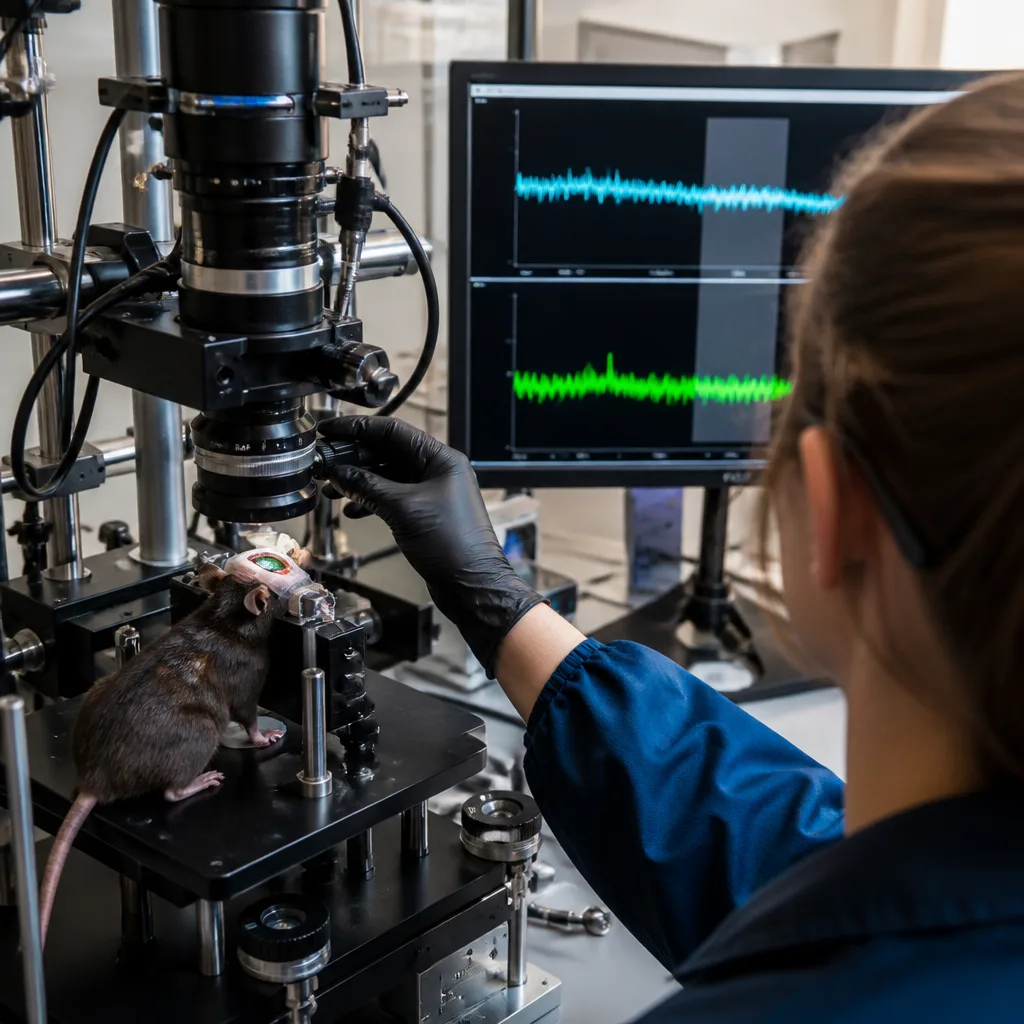
To probe energy dynamics in natural sleep, the team used an approach that kept the mouse skull transparent, sealed with a UV-curable resin, to allow prolonged optical access. Using wide-field fluorescence imaging they monitored three complementary signals:
- brain blood volume — an index of the vascular "fuel" arriving from circulation;
- neuronal ATP — the immediate molecule neurons use to power electrical and biochemical work;
- astrocytic pyruvate — a metabolite that links blood-derived glucose to cellular energy pathways.
Key findings and timing
During non-REM sleep the researchers observed the expected strong activity in the delta frequency band, together with subtler theta-band oscillations. Importantly, the team found that theta-band fluctuations predicted subsequent changes in brain blood volume by several seconds, implying that ongoing neuronal rhythms modulate vascular responses and thus metabolic supply.
By contrast, the transition into REM sleep revealed a different pattern. The study reports that measures of vascular supply increased around the period when REM emerges, while simultaneously the neuronal ATP signal fell — a paradoxical dissociation between supply and the molecule directly used by neurons.
"We were intrigued by this paradox, and wanted to look into the scientific basis behind why dreaming is somehow tiring." — Professor Ko Matsui, Tohoku University
What the results mean
The observations indicate that the sleeping brain does not simply switch off energetically. Instead, vascular delivery and local metabolic states can be decoupled: increased blood volume does not necessarily translate immediately into higher neuronal ATP. The involvement of astrocytic pyruvate measurements suggests the study probed intermediate metabolic steps between blood-borne glucose and neuronal energy supply.
Those temporal relationships — neuronal rhythms predicting vascular changes by seconds, and the rise in supply occurring alongside a fall in ATP — point to a nuanced, dynamic regulation of energy that depends on state and time. The work highlights how REM sleep, often equated with vivid dreaming and memory processing, places specific and complex demands on brain metabolism.
| Signal | Role measured | Observed change around REM |
|---|---|---|
| Brain blood volume | Vascular fuel supply | Increased |
| Neuronal ATP | Immediate neuronal energy | Decreased |
| Astrocytic pyruvate | Metabolic intermediary | Monitored (linking glucose to ATP) |
Context and consequences
Understanding how the brain balances supply and demand during sleep has implications for theories of why sleep is restorative and how memory consolidation or dreaming might be metabolically costly. The dissociation between blood flow and ATP challenges simple interpretations of imaging signals that equate increased haemodynamics with increased neuronal energy availability.
Further work will be required to determine whether similar dynamics occur in humans and how these patterns relate to cognitive functions attributed to REM sleep, such as memory processing and emotional regulation. For now, the study provides a clearer, experimentally grounded view of the metabolic choreography that accompanies one of the brain's most enigmatic states.